
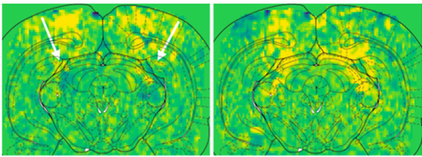
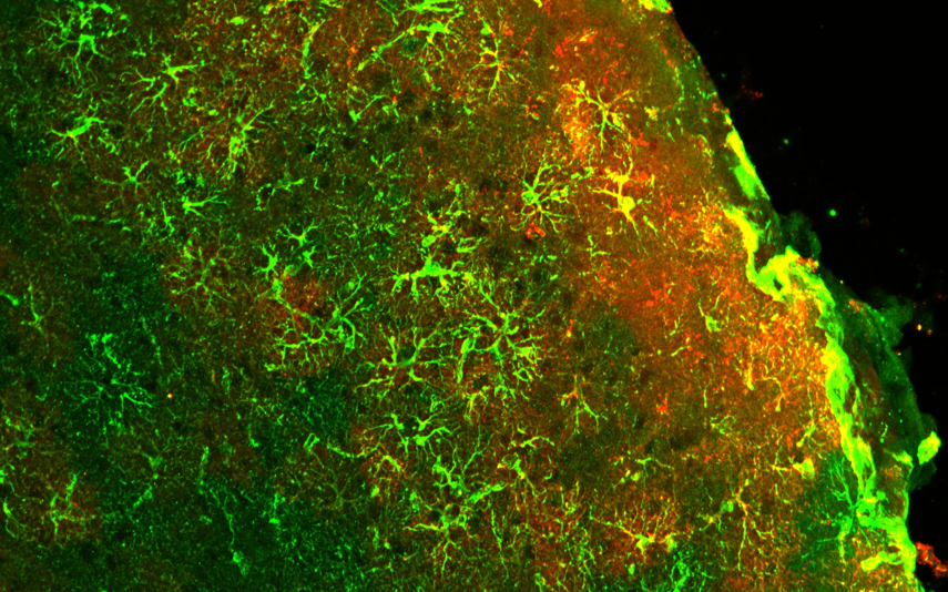
Cerebral hemodynamics are crucial for brain homeostasis and serve as a key proxy for brain activity. Although this process involves coordinated interaction between vessels, neurons, and glial cells, its dysregulation in neuroinflammation is not well understood. Here we used functional ultrasound imaging to investigate the impact of reactive glial cells, and in particular astrocytes, on cerebral blood volume changes during neuroinflammation. We found that reactive astrocytes increased the amplitude and propagation of vasomotion, a spontaneous oscillation of the cerebral blood volume around 0.1 Hz.
Astrocytes play a critical role in brain homeostasis and energetics, yet their exact contribution to neurovascular and neurometabolic coupling is not fully elucidated. In particular, how astrocytic activity participates in shaping the functional neuroimaging signals relying on these processes remains unclear. In this study, we used chemogenetic modulation of astrocytes in the primary visual cortex to investigate their influence on hemodynamic fluctuations using functional ultrasound imaging (fUSi), as well as glucose uptake using [18F]FDG-positron emission tomography (PET) in the same mice.
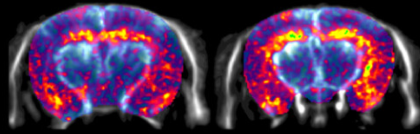
The 18 kDa translocator protein (TSPO) is used as a biomarker of neuroinflammation, but it may also play a direct role in the pathophysiology of neurological conditions with an inflammatory component such as Alzheimer’s disease. Here, we used functional ultrasound imaging (fUSi) to assess how TSPO knockout influences brain activity and connectivity in the 3xTgAD mouse model of AD.