
May 28, 2026
Brain activity is followed by changes in both cerebral blood flow and energy consumption. These relationships form the basis of several functional neuroimaging techniques: fMRI and functional ultrasound imaging rely on hemodynamic changes, while positron emission tomography (PET) with glucose analogue [18F]FDG measures glucose uptake. Both are commonly used as indirect windows onto brain function.
However, blood flow and glucose metabolism are not always strictly correlated, and the cellular mechanisms shaping these two signals are only partly understood. Astrocytes are particularly interesting in this context. Their processes interact closely with both synapses and blood vessels, and they have been implicated in the regulation of cerebral hemodynamics as well as in glucose metabolism. Yet these two aspects of astrocyte function have mostly been investigated separately.
In this study, we wanted to look at both responses in the same animals. We chemogenetically activated Gq signaling in astrocytes of the mouse visual cortex and combined functional ultrasound imaging (fUSi), to measure cerebral blood volume dynamics, with [18F]FDG-PET, to assess glucose uptake (on separate imaging days). The question was: could astrocyte activation affect these two functional imaging signals in parallel, or differently?
Using fUSi, we found two main effects of astrocytic activation.
At rest, there was no clear sustained increase or decrease in cerebral blood volume. Instead, astrocyte activation amplified spontaneous oscillations around 0.2 Hz, which we interpret as a relatively fast form of vasomotion (a spontaneous vasodilation/vasoconstriction cycle). This result was particularly interesting in light of our previous work, where we found that reactive astrocytes were involved in the amplification of vasomotion during neuroinflammation. Here, using a very different experimental approach, astrocytic Gq activation again increased the amplitude of an ongoing vasomotor process, and that, only in the transfected area (the visual cortex).
Importantly, these experiments were performed under medetomidine sedation, a physiological state associated with strong vasoconstriction and pronounced hemodynamic oscillations at relatively high frequencies. So the exact frequency observed here is probably closely linked to this experimental condition. Still, it provided an interesting setting to show that astrocytic activity can amplify ongoing vasomotion rather than simply alter mean blood volume.
Astrocyte activation also increased functional hyperemia, the local rise in cerebral blood volume induced by visual stimulation. In other words, astrocytic Gq signaling influenced both spontaneous and stimulus-evoked hemodynamic activity.
We then looked at glucose metabolism using [18F]FDG-PET in the same mice. Here, the result was different: astrocytic activation did not produce a significant increase in [18F]FDG uptake, even localized in the visual cortex. This was probably the most unexpected result, given the proposed role of astrocytes in cerebral glucose metabolism and previous work suggesting that astrocytic activity can contribute strongly to the FDG-PET signal.
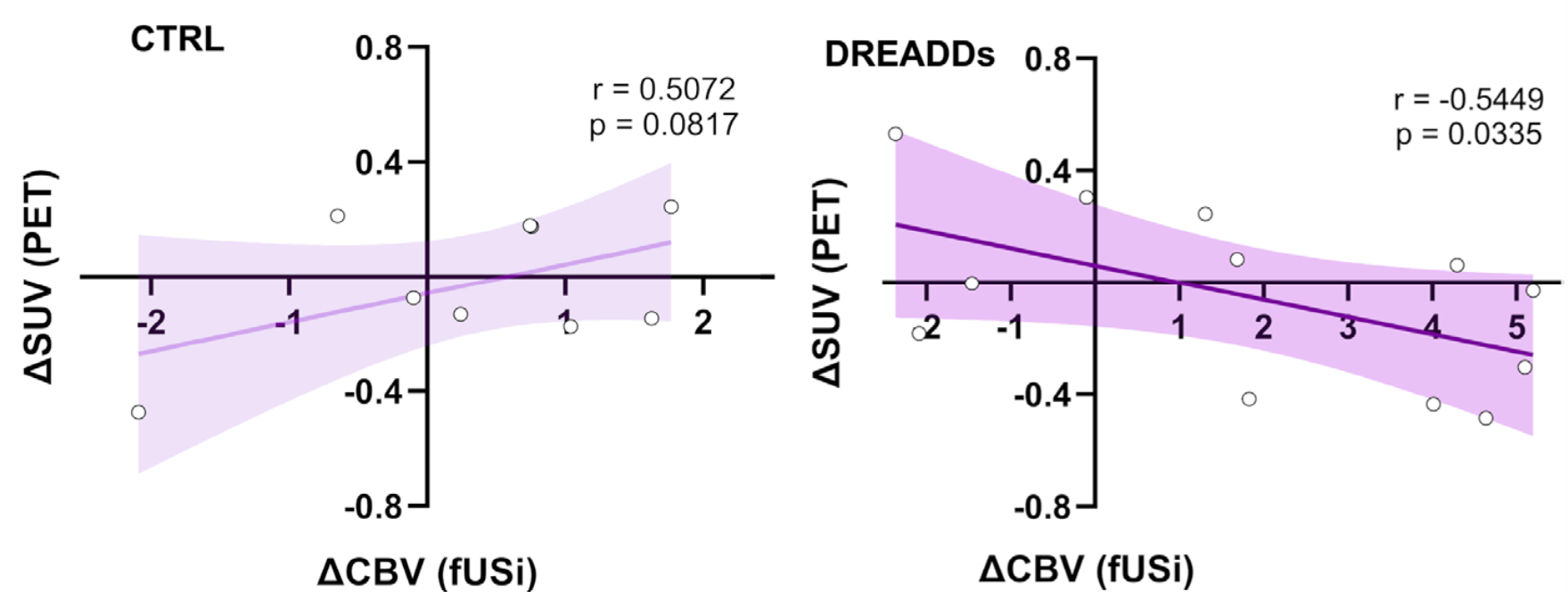
Furthermore, when we compared the two modalities across animals, we found a negative correlation between the change in functional hyperemia and the change in FDG uptake following astrocytic activation: animals showing the largest increase in hemodynamic response tended to show the largest decrease in FDG uptake.
This does not mean that astrocyte activation simply reduces glucose metabolism, as the average PET signal did not significantly change. But it does suggest that astrocytes can modify the relationship between vascular and metabolic responses.
Cerebral hemodynamics and glucose uptake are both widely used as indirect measures of brain activity, and they often evolve together. But they are not generated by exactly the same biological processes. Our results suggest that astrocytes may be one source of this dissociation. Why this happens remains speculative. One possibility discussed in the paper involves lactate metabolism. If astrocytic activation promotes glycolysis and lactate production, neurons could partly use lactate as an alternative energetic substrate, potentially reducing their need for direct glucose uptake. Lactate itself may also contribute to vascular responses. This could, at least conceptually, produce stronger hyperemia without a parallel increase in FDG uptake.
This is another reminder that functional neuroimaging signals are not simple readouts of neuronal activity. They also reflect the contribution of glial and vascular processes, and understanding these contributions may help us interpret why different imaging modalities sometimes tell slightly different stories.