
May 31, 2026
Alzheimer’s disease is usually discussed in terms of amyloid and tau accumulation or neuronal dysfunction/degeneration, but inflammation is also an important component of the pathology. One of the most widely used markers of this inflammatory response is the 18 kDa translocator protein, or TSPO, which can be measured in patients using PET imaging. But TSPO may be more than a passive biomarker, as emerging work suggests that TSPO directly participates to inflammation and cognitive alterations in Alzheimer's disease.
In this collaborative study with Millet Lab (Benjamin Tournier and Aurélien Badina) at the University of Geneva, we wanted to look at this question with a system-level scale: does TSPO also influence the organization of functional brain networks in Alzheimer’s disease? We used functional ultrasound imaging (fUSi) to record resting-state cerebral blood volume fluctuations in 16-month-old triple-transgenic 3xTgAD mice (a widely-used preclinical model that displays amyloid and tau burden along with inflammation, behavioral impairment and neuronal death), with or without genetic deletion of TSPO. Rather than starting from predefined regions, we used data-driven approaches to identify functional networks and characterize their dynamics.
Maybe not surprisingly, hippocampus emerged as the main region showing functional abnormalities. More interestingly, these alterations were not homogeneous.
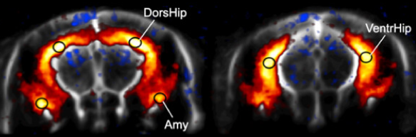
Over the full 10-minute resting-state acquisition, 3xTgAD mice showed stronger engagement of the ventral hippocampus within a bilateral hippocampal–amygdala network, together with increased local hemodynamic variance. This alteration was absent in 3xTgAD mice lacking the TSPO protein.
We then looked at faster network dynamics using co-activation pattern analysis, which identifies recurring spatial configurations of brain activity frame by frame. At this shorter, second-scale, we found another alteration: 3xTgAD mice showed abnormally few transitions from an activated to a deactivated state involving the dorsal hippocampus and retrosplenial cortex. Again, this alteration was prevented by TSPO knockout.
This shows that the same disease model can display different network abnormalities depending on both where and at what temporal scale we look. The ventral and dorsal hippocampus are functionally distinct regions, and here they also appeared to be differently affected by Alzheimer’s pathology, which may participate to distinct symptoms in the model.
Importantly, TSPO deletion normalized the functional network alterations but failed to reduce amyloid or phosphorylated tau levels. By contrast, astrocytic GFAP labeling was reduced, consistent with previous work showing that TSPO deletion limits astrocyte reactivity in this model. This further is in line with the idea that pathological protein burden is not the full story in Alzheimer's disease, and suggests that modulating inflammatory pathways can improve brain network function even without substantially reducing accumulation of amyloid and tau.