
May 27, 2025
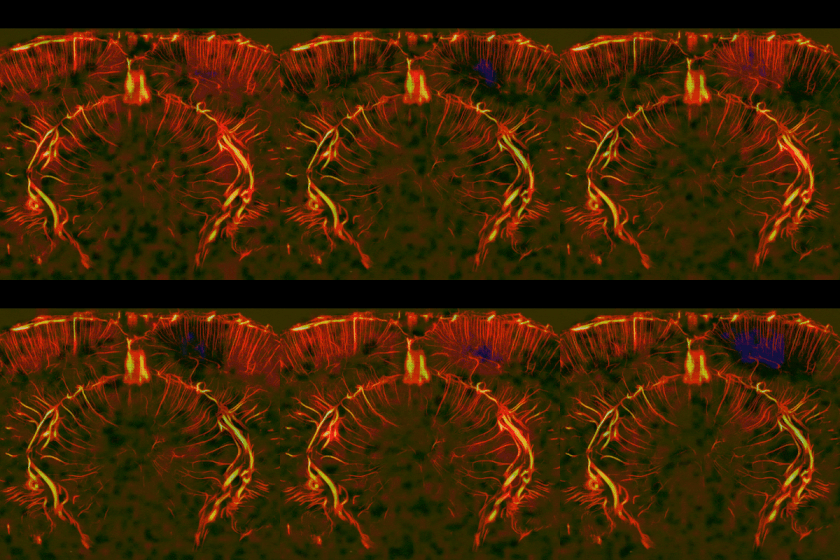
Brain activity is constantly followed by changes in blood flow, a process known as neurovascular coupling. This relationship is at the heart of functional neuroimaging: techniques such as fMRI or functional ultrasound rely on hemodynamic signals as an indirect window onto brain function.
But cerebral blood vessels are not simply passive reporters of neuronal activity. Their dynamics is actually shaped by the complex interaction between vascular cells, neurons and glia (non-neuronal cells). In this study, we asked what happens to these dynamics in the brain in the presence of a local inflammation. While I personally expected subtle changes, we instead encountered a rather striking phenomenon.
Using functional ultrasound imaging in a rat model of local cortical inflammation, we observed the emergence of large, almost sinusoidal oscillations in cerebral blood volume. This is unusual, and my initial interpretation was that there was probably some non-specific electronic artifact polluting our data. Turns out that this was a real biological effect. The signal had a period of roughly ten seconds, corresponding to a frequency around 0.1 Hz, the typical frequency of vasomotion, a spontaneous rhythmic contraction and dilation of blood vessels that is driven by intrinsic calcium oscillation in vascular smooth muscle cells.
The spatial organization of these oscillations was also quite interesting. When looking at the series of images, the changes in blood volume did not occur everywhere in the inflammed area at once. Instead, they travelled. We could follow waves moving from one side to the other side of the cortex, sometimes crossing several millimeters. While such effects have been reported using optic imaging techniques at the level of the superficial pial vessels that rely on top of the cortex, here thanks to functional ultrasound we could see that the waves involved neighbouring penetrating vessels and could span the full cortical thickness. Some waves propagated even farther, linking cortical and subcortical structures. Inflammation increased both the occurrence of these travelling events and the distances over which they propagated, meaning that vasomotion waves are already present in normal conditions, but they can be drastically enhanced in certain pathological states.
This was perhaps the part that interested us most.
Neuroinflammation profoundly changes the state of glial cells, in particular microglia and astrocytes. Astrocytes are especially well positioned to influence vascular dynamics: their endfeet surround much of the cerebral vasculature, placing them directly at the interface between neural tissue and blood vessels. Under normal conditions, they are already known to participate in the regulation of cerebral perfusion and neurovascular coupling.
We therefore asked whether reactive glia could contribute to the amplification of the vasomotor waves that we were observing. The regions and animals displaying stronger ~0.1 Hz oscillations also showed stronger changes in microglial morphology. But the experiment pointed toward astrocytes more directly. When we induced astrocytic lesions using two different pharmacological approaches, the dramatic increase in vasomotion produced by inflammation was strongly reduced or essentially prevented. And it was not only the amplitude of the oscillations that was normalized. The abnormal long-distance travelling-wave patterns and the associated reorganization of hemodynamic activity were also largely lost.
This suggests that reactive astrocytes are important amplifiers of inflammatory vasomotion. We do not yet know the precise molecular mechanism, and the interaction is probably not a simple one-way pathway: microglia, astrocytes, vascular smooth-muscle cells and neurons may all participate. But our results place astrocytes much closer to the centre of the phenomenon than one might expect from a purely neuron-centred interpretation of functional imaging.
A ~0.1 Hz fluctuation sits directly in the frequency range commonly investigated in resting-state functional imaging. If inflammation can strongly amplify vasomotion and organize it into waves travelling across distant brain regions, then some of the correlations measured in functional imaging may reflect more than coordinated neuronal activity. They may also contain information about the state of the glial systems.
My (rather optimistic) take is that this does not make hemodynamic imaging less useful. It suggests that these signals may carry a lot of biological information that we are only beginning to decode, and this goes beyond local correlation with neuronal activity. In pathological conditions, pronounced ~0.1 Hz oscillations or travelling vascular waves could potentially tell us something about glial reactivity itself, and overall inflammation.